Your Health..Our Mission..
For Emergency Call +91-9167870733


+91- 9158-80-6000
+91- 9168-20-6000
+91- 9168-20-6000
Opp Everest World, Gate No.
2, Kolshet Road.
2, Kolshet Road.
MONDAY TO SATURDAY
07:00 AM - 10:00 PM
SUNDAY 07:00 AM - 04:00 PM
07:00 AM - 10:00 PM
SUNDAY 07:00 AM - 04:00 PM
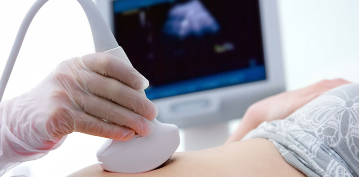
Ultrasonography
Ultrasonography, also known as ultrasound imaging or diagnostic medical sonography, is a non-invasive medical imaging technique that uses high-frequency sound waves to create images of the internal structures of the body. It is widely used in various medical specialties, including radiology, obstetrics and gynecology, cardiology, and many others.
Sonography Timing
Morning 10.00 AM to 12.00 PM (Male Radiologist)
Afternoon 02:30 PM to 03:30 PM (Female Radiologist)
Here's how ultrasonography works and some key features:
- Sound wave generation: During an ultrasonography exam, a handheld device called a transducer is used. The transducer emits high-frequency sound waves into the body's tissues.
- Sound wave propagation: The sound waves travel through the body and encounter different tissues and organs. As they encounter boundaries between tissues of varying densities, some of the sound waves are reflected back to the transducer.
- Echo formation: The reflected sound waves, known as echoes, are detected by the transducer. The transducer converts these echoes into electrical signals.
- Image formation: The electrical signals are processed by a computer, which creates real-time images based on the timing and strength of the echoes. These images are displayed on a monitor and can be interpreted by a healthcare professional.
- Non-invasive and safe: Ultrasonography is non-invasive, meaning it does not involve any surgical incisions or radiation exposure. It is considered safe and is generally well-tolerated by patients. It can be used in patients of all ages, including infants and pregnant women.
- Versatile imaging modality: Ultrasonography can be used to visualize a wide range of organs and tissues in the body. It is commonly used to examine the abdomen, pelvis, heart, blood vessels, muscles, tendons, and superficial structures like the thyroid gland and breasts.
- Real-time imaging: One of the major advantages of ultrasonography is the ability to produce real-time images. This allows healthcare professionals to visualize dynamic processes, such as the movement of the heart, blood flow, or the motion of a fetus during pregnancy.
- Guided procedures: Ultrasonography is often used as a guide for various medical procedures. For example, it can assist in needle biopsies, fluid aspiration, or the placement of catheters or drainage tubes.
Ultrasonography has numerous clinical applications and is a valuable tool in medical diagnosis and management. It provides detailed images of internal structures in real time, aiding in the detection of abnormalities, guiding interventions, and monitoring treatment progress. It is a widely available and widely used imaging modality due to its safety, versatility, and real-time capabilities.
©2023 All Rights Reserved by Kolshet Diagnostics.