Your Health..Our Mission..
For Emergency Call +91-9167870733


+91- 9158-80-6000
+91- 9168-20-6000
+91- 9168-20-6000
Opp Everest World, Gate No.
2, Kolshet Road.
2, Kolshet Road.
MONDAY TO SATURDAY
07:00 AM - 10:00 PM
SUNDAY 07:00 AM - 04:00 PM
07:00 AM - 10:00 PM
SUNDAY 07:00 AM - 04:00 PM
2D Echo
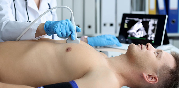
A 2D echocardiogram, commonly referred to as a 2D echo, is a non-invasive imaging test used to visualize the structure and function of the heart using ultrasound technology. It provides a two-dimensional real-time moving image of the heart, allowing healthcare professionals to assess the heart's chambers, valves, and overall cardiac function.
Here's how a 2D echo works and its key features:
- Ultrasound imaging: A 2D echo utilizes ultrasound waves to generate images of the heart. A transducer, a handheld device, is placed on the patient's chest and emits high-frequency sound waves. These sound waves bounce off the heart's structures and are detected by the transducer, which converts them into electrical signals.
- Real-time moving images: The electrical signals are processed by a computer, which creates a two-dimensional moving image of the heart on a monitor. This real-time imaging allows healthcare professionals to observe the heart's movement, including the contracting and relaxing of the chambers, as well as the opening and closing of the heart valves.
- Structural assessment: A 2D echo provides detailed information about the heart's structure. It allows visualization of the heart's walls, chambers, valves, and major blood vessels. This enables the detection of any structural abnormalities, such as thickening of the heart muscle (hypertrophy), presence of tumors, or congenital heart defects.
- Assessment of heart function: The 2D echo provides valuable information about the heart's function. It allows the evaluation of the heart's pumping ability, the measurement of the ejection fraction (the percentage of blood pumped out with each heartbeat), and the assessment of the heart valves' performance, including any leakage or narrowing.
- Doppler imaging: In addition to the 2D images, a 2D echo can incorporate Doppler imaging. Doppler ultrasound measures the speed and direction of blood flow within the heart and blood vessels. This helps in the assessment of blood flow patterns, identification of abnormal flow, and detection of valve regurgitation or stenosis.
- Non-invasive and safe: A 2D echo is a non-invasive procedure, meaning it does not involve any surgical incisions or radiation exposure. It is considered safe and well-tolerated, with no known risks or side effects associated with the test. It can be performed on patients of all ages, including infants and pregnant women.
A 2D echo is a valuable diagnostic tool used in cardiology to assess various heart conditions, such as heart valve disorders, heart failure, congenital heart defects, and abnormal heart rhythms. It provides detailed information about the heart's structure and function, aiding in accurate diagnosis, treatment planning, and monitoring of cardiac conditions.
©2023 All Rights Reserved by Kolshet Diagnostics.